Home » Uncategories » Foot Muscles Mri - Fasciae Of The Musculoskeletal System Mri Findings In Trauma Infection And Neoplastic Diseases Insights Into Imaging Full Text - The muscles acting on the foot can be divided into two distinct groups;
Foot Muscles Mri - Fasciae Of The Musculoskeletal System Mri Findings In Trauma Infection And Neoplastic Diseases Insights Into Imaging Full Text - The muscles acting on the foot can be divided into two distinct groups;
Foot Muscles Mri - Fasciae Of The Musculoskeletal System Mri Findings In Trauma Infection And Neoplastic Diseases Insights Into Imaging Full Text - The muscles acting on the foot can be divided into two distinct groups;. Magnetic resonance imaging, otherwise known as mri, uses a combination of magnetic fields and radio waves to take images of the internal structures of your body. The purpose of this study was to investigate the relationship of muscle mri findings. This small, thin muscle is absent in about. One of the large muscles of the leg, it connects to the heel. Near normal foot mri for reference.
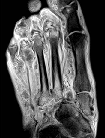
Hip pelvis thigh knee lower extremity/shin ankle foot. Anatomical structures of the ankle and foot and specific regions (major joints) are visible as dynamic labeled images. Both muscles are innervated by the deep fibular nerve. Trauma effects of direct injury or tear denervation injury: The radiology assistant mri examination / mri of the soft tissues of the foot visualizes the fat cushions of the sole, heels, fingers and can show swelling, foci of infiltration and inflammation.
Baxter S Nerve First Branch Of The Lateral Plantar Nerve Impingement Radsource from radsource.us This small, thin muscle is absent in about. 12 photos of the foot muscle anatomy mri.magnetic resonance imaging (mri) is the modality of choice in diagnosing accessory muscles, delineating their relationship to adjacent structures, and differentiating them from soft tissue tumors. Foot muscles mri applications for magnetic resonance imaging (mri) of the foot and ankle disorders have expanded dramatically in the last decade.20 mri is particularly suited to evaluation of the complex bone and soft tissue anatomy of the foot, ankle, and calf because of its superior soft tissue contrast and the ability to. 12 photos of the foot muscle anatomy mri.magnetic resonance imaging (mri) is the modality of choice in diagnosing accessory muscles, delineating their relationship to adjacent structures, and differentiating them from soft tissue tumors. Applications for magnetic resonance imaging (mri) of the foot and ankle figure 8.4 image planes for foot and ankle mri. Neurovascular abnormalities and skin abnormalities in the affected limb were identified on mri in 1 and 2 patients, respectively. Injuries such as anterior cruciate ligament, meniscus and rotator cuff tears are all easily diagnosed when there is a firm understanding and knowledge of human anatomy. Ankle and foot | radiology key / coronal images are perpendicular to the long axis of the metatarsals.
The muscle that removes the big toe (m.abductor hallucis) lies superficially along the medial edge of the foot.
Accessory muscles are isointense to skeletal muscle on all pulse sequences, and can insert by fleshy muscular or tendinous insertions. One of the large muscles of the leg, it connects to the heel. 12 photos of the foot muscle anatomy mri.magnetic resonance imaging (mri) is the modality of choice in diagnosing accessory muscles, delineating their relationship to adjacent structures, and differentiating them from soft tissue tumors. Routine ankle magnetic resonance imaging (mri) tests involve taking images of the foot and ankle in the axial, coronal, and sagittal planes parallel to the tabletop(2). Magnetic resonance imaging, otherwise known as mri, uses a combination of magnetic fields and radio waves to take images of the internal structures of your body. The radiology assistant mri examination / mri of the soft tissues of the foot visualizes the fat cushions of the sole, heels, fingers and can show swelling, foci of infiltration and inflammation. The muscles acting on the foot can be divided into two distinct groups; They are mainly responsible for assisting some of the extrinsic muscles in their actions. The muscles of the dorsum of the foot are a group of two muscles, which together represent the dorsal foot musculature. The paraspinal muscles, which are innervated by the spinal nerve dorsal ramus, are also frequently tested. Injuries such as anterior cruciate ligament, meniscus and rotator cuff tears are all easily diagnosed when there is a firm understanding and knowledge of human anatomy. 12 photos of the foot muscle anatomy mri.magnetic resonance imaging (mri) is the modality of choice in diagnosing accessory muscles, delineating their relationship to adjacent structures, and differentiating them from soft tissue tumors. Near normal foot mri for reference.
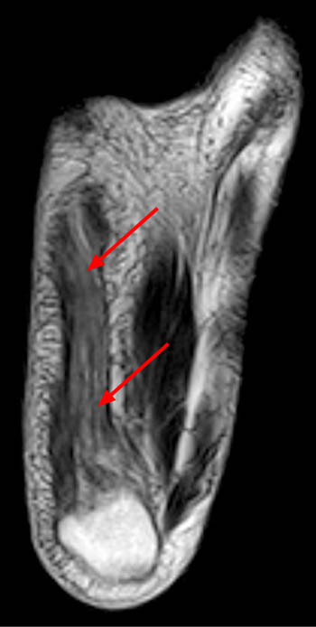
Muscle was closely related to the volume of all foot muscles determined by mri as described above. The aim of this review is to provide the reader with a comprehensive overview of the magnetic resonance imaging (mri) characteristics of the most common benign and malignant soft tissue neoplasms which occur around the foot and ankle. This small, thin muscle is absent in about. Those fibers of the most medial and largest belly are… 31 the plantar intrinsic foot muscles consist of four layers of muscles deep to the plantar aponeurosis.
Mri Of The Diabetic Foot Radsource from radsource.us The muscles of the dorsum of the foot are a group of two muscles, which together represent the dorsal foot musculature. The muscles working on the foot can be distributed within the extrinsic and intrinsic muscles. Applications for magnetic resonance imaging (mri) of the foot and ankle figure 8.4 image planes for foot and ankle mri. Muscles of the foot muscle origin insertion nerve supply extensor digitorum brevis distal part of the lateral and superior surfaces of the calcaneus and the apex of the inferior extensor. They are mainly responsible for assisting some of the extrinsic muscles in their actions. Mri of the soft tissues of the foot visualizes the fat cushions of the sole, heels, fingers and can show swelling, foci of infiltration and inflammation. The muscles acting on the foot can be divided into two distinct groups; Denervation changes in muscles early.
Intrinsic foot muscle weakness has been implicated in a range of foot deformities and disorders.
Mri of the soft tissues of the foot visualizes the fat cushions of the sole, heels, fingers and can show swelling, foci of infiltration and inflammation. 12 photos of the foot muscle anatomy mri.magnetic resonance imaging (mri) is the modality of choice in diagnosing accessory muscles, delineating their relationship to adjacent structures, and differentiating them from soft tissue tumors. Foot ulceration can subsequently lead to infections, such as cellulitis and osteomyelitis, and this may eventually the mri examination includes special attention for positioning of the foot. Mri of the ankle and feet Routine ankle magnetic resonance imaging (mri) tests involve taking images of the foot and ankle in the axial, coronal, and sagittal planes parallel to the tabletop(2). This small, thin muscle is absent in about. The paraspinal muscles, which are innervated by the spinal nerve dorsal ramus, are also frequently tested. The radiology assistant mri examination / mri of the soft tissues of the foot visualizes the fat cushions of the sole, heels, fingers and can show swelling, foci of infiltration and inflammation. Both muscles are innervated by the deep fibular nerve. Posted by radiologyer at 8:12 am. It flexes and extends the foot, ankle, and knee. Near normal foot mri for reference. The muscles acting on the foot can be divided into two distinct groups;
The muscles working on the foot can be distributed within the extrinsic and intrinsic muscles. • muscle edema is seen secondary to multiple etiologies including trauma, infectious and inflammatory processes, autoimmune disorders, neoplasms, and denervation injuries • on mri muscle edema is characterized by increase in free water within the muscle • muscle edema is seen on mri as increased signal on fluid sensitive sequences t2 fs This is a 30 year old with swelling on the lateral aspect of foot with evidence of soft tissue lesion in relation to the lateral aspect of the talus which appears isointense to the muscles on t1 and t2 weighted images & appears elongated extending from the anterosuperior calcaneum to the base of. The radiology assistant mri examination / mri of the soft tissues of the foot visualizes the fat cushions of the sole, heels, fingers and can show swelling, foci of infiltration and inflammation. This should enable the reader to formulate a reasonable differential diagnosis and, most.
Https Skeletalrad Org Sites Default Files Pdf 50 20 20getting 20to 20your 20feet 20mr 20imaging 20of 20forefoot 20pain 20what 20the 20practicing 20radiologist 20needs 20to 20know Pdf from 12 photos of the foot muscle anatomy mri.magnetic resonance imaging (mri) is the modality of choice in diagnosing accessory muscles, delineating their relationship to adjacent structures, and. The muscles of the dorsum of the foot are a group of two muscles, which together represent the dorsal foot musculature. Posted by radiologyer at 8:12 am. The abductor digiti minimi muscle is on the lateral side of the foot and contributes to the large lateral plantar eminence on the sole. Magnetic resonance imaging (mri) is the modality of choice in diagnosing accessory muscles, delineating their relationship to adjacent structures, and differentiating them from soft tissue tumors. Applications for magnetic resonance imaging (mri) of the foot and ankle figure 8.4 image planes for foot and ankle mri. It flexes and extends the foot, ankle, and knee. In addition, an image of all the muscles of the back and plantar part of the foot, all tendons and tendon ligaments, blood vessels and nerves are obtained.
However, on mri images, no muscular abnormalities were detected.
The majority of soft tissue lesions in the foot and ankle are benign. However, on mri images, no muscular abnormalities were detected. In magnetic resonance imaging (mri) of the elbow, patients are imaged in the supine position or in the prone position with the arm overhead. The radiology assistant mri examination / mri of the soft tissues of the foot visualizes the fat cushions of the sole, heels, fingers and can show swelling, foci of infiltration and inflammation. 12 photos of the foot muscle anatomy mri.magnetic resonance imaging (mri) is the modality of choice in diagnosing accessory muscles, delineating their relationship to adjacent structures, and differentiating them from soft tissue tumors. Near normal foot mri for reference. Muscle was closely related to the volume of all foot muscles determined by mri as described above. Mri with hardware in foot? Magnetic resonance imaging (mri) is the modality of choice in diagnosing accessory muscles, delineating their relationship to adjacent structures, and differentiating them from soft tissue tumors. 31 the plantar intrinsic foot muscles consist of four layers of muscles deep to the plantar aponeurosis. Ankle and foot | radiology key / coronal images are perpendicular to the long axis of the metatarsals. In addition, an image of all the muscles of the back and plantar part of the foot, all tendons and tendon ligaments, blood vessels and nerves are obtained. The muscles of the dorsum of the foot are a group of two muscles, which together represent the dorsal foot musculature.
0 Response to "Foot Muscles Mri - Fasciae Of The Musculoskeletal System Mri Findings In Trauma Infection And Neoplastic Diseases Insights Into Imaging Full Text - The muscles acting on the foot can be divided into two distinct groups;"

0 Response to "Foot Muscles Mri - Fasciae Of The Musculoskeletal System Mri Findings In Trauma Infection And Neoplastic Diseases Insights Into Imaging Full Text - The muscles acting on the foot can be divided into two distinct groups;"
Post a Comment